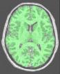
Compute a binary mask of the brain from a bias corrected T1-weighted image
The basic principles of this procedure are described in Ana Brain Mask from T1 MRI and Correction Brain Mask from T1 MRI. Moreover, the log files are full of information...
In this low level access, you can tune some parameters to overcome difficult cases. If it is still a failure, your last hope is in using the commandline procedure, which gives access to much more parameters. An interesting alternative, however, is manual drawing, at least to clean up problematic areas... Semi-manual drawing may be especially wellcome when a lesion is disturbing the segmentation process.
For more information:
Robust brain segmentation using histogram scale-space analysis and mathematical morphology, J.-F. Mangin, O. Coulon, and V. Frouin MICCAI, MIT, LNCS-1496, Springer Verlag 1230-1241, 1998
You can also try the commandline
VipGetBrain -help
mri_corrected: T1 MRI Bias Corrected ( input )
histo_analysis: Histo Analysis ( input )
Grey/white statistics
mode: Choice ( input )
Commissure_coordinates: Commissure coordinates ( optional, input )
brain_mask: T1 Brain Mask ( output )
binary mask
regularization: Boolean ( input )during binarization
erosion_size: Float ( input )to get brain seed
layer: Choice ( input )
first_slice: Integer ( input )slices to erase at top
last_slice: Integer ( input )slices to erase at bottom
lesion_mask: Lesion Mask ( optional, input )binary mask (option)
Toolbox : Morphologist
User level : 2
Identifier :
VipGetBrainSupported file formats :
mri_corrected :GIS image, VIDA image, NIFTI-1 image, MINC image, gz compressed MINC image, DICOM image, TIFF image, XBM image, PBM image, PGM image, BMP image, XPM image, PPM image, gz compressed NIFTI-1 image, TIFF(.tif) image, ECAT i image, PNG image, JPEG image, MNG image, GIF image, SPM image, ECAT v imagehisto_analysis :Histo AnalysisCommissure_coordinates :Commissure coordinatesbrain_mask :GIS image, VIDA image, NIFTI-1 image, MINC image, TIFF image, XBM image, PBM image, PGM image, BMP image, XPM image, PPM image, gz compressed NIFTI-1 image, ECAT i image, PNG image, JPEG image, MNG image, GIF image, SPM image, ECAT v imagelesion_mask :GIS image, VIDA image, NIFTI-1 image, MINC image, gz compressed MINC image, DICOM image, TIFF image, XBM image, PBM image, PGM image, BMP image, XPM image, PPM image, gz compressed NIFTI-1 image, TIFF(.tif) image, ECAT i image, PNG image, JPEG image, MNG image, GIF image, SPM image, ECAT v image

