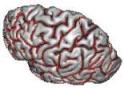

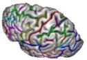
Visualisation of cortical fold graphs, with or without names
Ce traitement chargera également le maillage de l'hémisphère associé, et si vous le souhaitez l'IRM corrigée du biais.
graph: Cortical folds graph ( input )
nomenclature: Nomenclature ( optional, input )The hierarchy of names providing the color code
white_mesh: Hemisphere White Mesh ( optional, input )
hemi_mesh: Hemisphere Mesh ( optional, input )
load_MRI: Choice ( input )
two_windows: Choice ( input )
mri_corrected: T1 MRI Bias Corrected ( optional, input )
Toolbox : Morphologist
User level : 0
Identifier :
AnatomistShowFoldGraphFile name :
brainvisa/toolboxes/morphologist/processes/viewers/Sulci/AnatomistShowFoldGraph.pySupported file formats :
graph :Graph and datanomenclature :Hierarchywhite_mesh :TRI mesh, Z compressed TRI mesh, PLY mesh, gz compressed TRI mesh, MESH mesh, Z compressed MESH mesh, gz compressed MESH mesh, gz compressed PLY mesh, Z compressed PLY mesh, GIFTI file, Z compressed GIFTI file, MNI OBJ mesh, gz compressed GIFTI file, Z compressed MNI OBJ mesh, gz compressed MNI OBJ meshhemi_mesh :TRI mesh, Z compressed TRI mesh, PLY mesh, gz compressed TRI mesh, MESH mesh, Z compressed MESH mesh, gz compressed MESH mesh, gz compressed PLY mesh, Z compressed PLY mesh, GIFTI file, Z compressed GIFTI file, MNI OBJ mesh, gz compressed GIFTI file, Z compressed MNI OBJ mesh, gz compressed MNI OBJ meshmri_corrected :GIS image, Z compressed GIS image, gz compressed GIS image, VIDA image, NIFTI-1 image, gz compressed ECAT i image, MINC image, gz compressed MINC image, DICOM image, TIFF image, XBM image, PBM image, PGM image, BMP image, XPM image, PPM image, gz compressed NIFTI-1 image, TIFF(.tif) image, gz compressed VIDA image, Z compressed VIDA image, Z compressed ECAT i image, gz compressed ECAT v image, ECAT i image, Z compressed ECAT v image, PNG image, JPEG image, MNG image, GIF image, Z compressed SPM image, SPM image, gz compressed SPM image, ECAT v image